 |
| About Bioline | All Journals | Testimonials | Membership | News |
|
||||||
|
||||||
Annals of African Medicine, Vol. 4, No. 3, 2005, pp. 136-138 Case Report FETUS PAPYRACEOUS: A CASE REPORT L. R. Airede and Y. Ahmed Department of Obstetrics and Gynaecology,
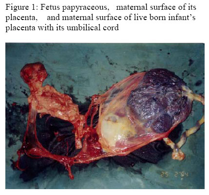
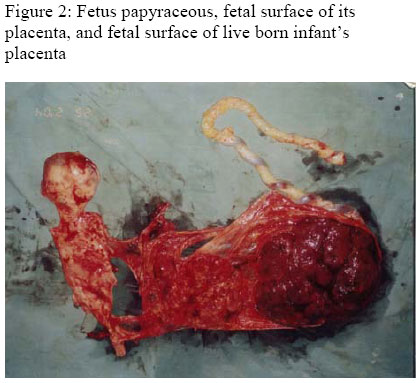
UsmanuDanfodiyoUniversity Teaching Hospital, Sokoto, Nigeria Code Number: am05034 Abstract Fetus papyraceous, the compressed remains of a dead twin retained in utero after its intrauterine death in the second trimester, is an uncommon finding. It is not usually associated with adverse physical effects on the mother or surviving twin. A case of fetus papyraceous which was discovered at UsmanuDanfodiyoUniversity teaching hospital, Sokoto, Nigeria during routine examination of the placenta and membranes after spontaneous vaginal delivery of a low birth weight, but otherwise healthy infant, to a primigravid mother is presented. No adverse effects on the mother were recorded. This is apparently the first reported case from Nigeria despite the high twinning rate in the country. Key words:Fetus papyraceous Résumé Foetus papyracé, les restes comprimés d'un jumeau mort maintenu dans l'utérus après sa mort intra-utérine dans le deuxième trimestre, est un résultat rare. Il n'est pas habituellement associé aux effets physiques défavorables sur la mère ou en jumeau survivant. Un cas du foetus papyracé qui a été découvert à l'hôpital d'enseignement de l’Université d'Usmanu Danfodiyo, Sokoto, au Nigéria, pendant l'examen courant du placenta et les membranes après l'accouchement vaginal spontanée d'une naissance de bas poids, mais autrement un nourrisson en bonne santé, à une mère primigeste est présenté. Aucun effet nuisible sur la mère n'a été enregistré. C'est apparemment le premier cas rapporté du Nigéria en dépit du taux élevé de gémellité dans le pays. Mots clés : Foetus papyraceous Introduction Fetus papyraceous or compressus is the compressed, mummified, parchment-like remains of a dead twin which is retained in-utero after intrauterine death in the second trimester.1-3 It is usually discovered among the placenta and membranes of its well-developed twin.2 The cause is thought to be death of one twin, amniotic fluid loss, or reabsorption and compression of the dead fetus.3 The incidence of fetus papyraceous has been reported at 1 in 17,000 to 20,000 pregnancies.3 Despite the high incidence of twinning in Nigeria, from which the highest incidence of twinning (53.8 per one thousand deliveries) is reported,4 fetus papyraceous has not been reported. Case report A 20 year old Nigerian primigravida was referred to the UsmanuDanfodiyoUniversity teaching hospital (UDUTH), Sokoto at a gestational age of 39 weeks, for delivery. She had booked for antenatal care at a primary health care center at 16 weeks gestation and regularly attended the clinic. Her packed cell volume at booking was 25%, sickling test was negative and her blood group was O, Rhesus positive. Urinalysis was negative for sugar and protein. Ultrasound scan at booking revealed a singleton, active fetus with a biparietal diameter of 35mm (gestational age 16 weeks), anterior placenta and adequate liquor volume. She was placed on ferrous sulphate tablets 200mg three times daily, folic acid tablets 5mg daily, and pyrimethamine tablets 25mg weekly for the duration of pregnancy. However, further investigation of her anaemia, or repeat of packed cell volume measurement was not done otherwise uneventful. She was a twin. She presented with a history of labour pains of seven hours duration. Clinical examination revealed a healthy, young woman, who was not pale and had no peripheral edema. Her pulse rate was 84 beats per minute, regular and of good volume, blood pressure was 100/70mm Hg, and her chest was clinically clear. Abdominal examination revealed an enlarged uterus with a symphysiofundal height of 35cm. There was a singleton fetus in longitudinal lie, cephalic presentation and right occipitoanterior position. The fetal heart rate was 140 per minute and regular. Vaginal examination revealed a normal vulva and vagina. The cervix was fully effaced, and the os was 5cm dilated. The fetal station was 0, the fetal membranes were intact, and the maternal pelvis was adjudged adequate for vaginal delivery. She was admitted into the labour ward and artificial rupture of the membranes was done. An intravenous access was obtained and blood obtained for packed cell volume and grouping and cross matching of 2 units of whole blood. Her packed cell volume was 32%. Five percent (5%) dextrose in normal saline was infused at a rate of 1litre 8-hourly. Two hours later, she had spontaneous vaginal delivery of a live female infant with Apgar scores of 6 and 8, at 1 and 5 minutes respectively. The infant’s birth weight was 2.15kg, she had no congenital abnormalities, and gestational age assessed by the Dubowitz et al criteria,5 was 39 weeks. The placenta was delivered by controlled cord traction and postpartum blood loss was 200mls. Examination of the placenta revealed one healthy looking placenta weighing 370g, and contiguous to it, a pale, atrophic placenta attached by a rudimentary cord to a fetus papyraceous. The fetus papyraceous and its placenta both weighed 150g. There were obvious musculoskeletal abnormalities of the fetus papyraceous (Figures 1 and 2). The baby was given 1 mg of Vitamin K1 intramuscularly 2 hours after delivery as was routine at UDUTH, and early feeding was instituted. Both mother and baby did well post partum and were discharged after 48 hours of observation. Discussion Despite the high rates of twinning reported from all parts of Nigeria, 4, 6 fetus papyraceous has not been more commonly reported from Nigeria. This may be as a result of the low number of births that take place in health facilities in Nigeria, or the thoroughness (or lack of it) with which the placenta is examined after delivery. A fetus papyraceous may be viewed with consternation by non-medical personnel if discovered after a home delivery. Prior to the use of ultrasound, the diagnosis of fetus papyraceous could only be made after delivery of the surviving twin. The advent of real time ultrasound permits the diagnosis of multiple gestation as early as four weeks after conception using the intravaginal probe.3 The subsequent demise of one fetus could then be diagnosed before delivery. In this case, however, mid trimester ultrasound at 16 weeks failed to diagnose twin gestation. No specific complications of fetus papyraceous in either mother or the surviving twin are reported in the literature. It would be expected that, as in singleton pregnancies, in utero retention of a fetus after its death would trigger disseminated intravascular coagulation in the mother. In twin pregnancies, however, the incidence of this complication is very low7. In the study by Malinowski et al, it was also found that none of the live born fetuses had evidence of hematological abnormalities.8 In the case presented, the surviving twin was low birth weight and small for gestational age, weighing only 2.15kg at birth despite the estimated gestational age of 39 weeks. It would not be possible however to ascribe the low birth weight of the survivor to the fetus papyraceous because the mother had anaemia at 16 weeks of pregnancy, which was not investigated at the referring clinic, but could have resulted in low birth weight.9 The musculoskeletal abnormalities found in the fetus papyraceous itself may have resulted from prolonged maceration and compression in utero. Further information which an autopsy may have provided was not available. In conclusion, a case of fetus papyraceous with no confirmed adverse effect on the mother or surviving twin is described. The cause of intrauterine death of the fetus could not be ascertained. In order that the diagnosis of multiple pregnancy is not missed during ultrasound examination, it is recommended that those who practice obstetric ultrasound obtain a high degree of proficiency by better training, and maintain a high index of suspicion. The use of modern ultrasound machines with good resolution is also advocated. It is also a sine qua non of good management of labour that the placenta and membranes are thoroughly inspected. This will permit the diagnosis of abnormalities of the placenta and membranes, and of fetus papyraceous as occurred in this case. References
Copyright 2005 - Annals of African Medicine The following images related to this document are available:Photo images[am05034f1.jpg] [am05034f2.jpg] |
| |||||||||