 |
| About Bioline | All Journals | Testimonials | Membership | News |
|
||||||
|
||||||
Letters to the Editor Linear cutaneous lupus erythematosus Antonis A. Kousoulis1,2, Ilianna Galli-Vareia1, Foteini Kousathana1,2, Ioanna-Maria Athanasopoulou1 History of Medicine Department, 1Medical School, University of
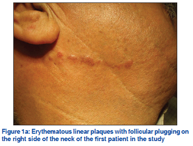
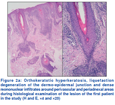
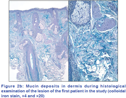
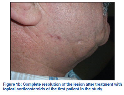
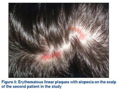
Athens, Athens, 2Society of Junior Doctors, Athens, Greece Code Number: dv11217 Sir, Linear lesions following the lines of Blaschko of discoid lupus erythematosus (DLE), subacute cutaneous lupus erythematosus or tumid lupus erythematosus are called linear cutaneous lupus erythematosus (LCLE). LCLE is a highly unusual variant of cutaneous lupus. It occurs mainly in children and young adults with a similar incidence in both sexes, and photosensitivity is less frequent. Lesions appear as linear unilateral erythematous plaques and predominate in the face; however, neck, trunk or the extremities may also be affected. In the majority of the cases, lesions are restricted to one anatomical site, and only one 8-year old boy showed widespread DLE to our best knowledge in literature search.[1] We report two cases of adult patients (≥15 years old) with LCLE. To our knowledge, there are only 10 previous reports of LCLE in adults. A 64-year-old male with a previous medical history significant for ankylosing spondylitis was referred because of a 7 month history of a persisting asymptomatic linear erythematus lesion on the right side of his neck. No history of preceding trauma or exposure to sunlight was present. The lesion showed follicular plugging [Figure 1a]. The results of routine laboratory tests were normal and antinuclear antibodies (ANAs) were absent. Histological examination revealed orthokeratotic hyperkeratosis with keratotic plaques and liquefaction degeneration of the dermo-epidermal junction. Dense mononuclear infiltrates around perivascular and periadnexal areas were seen in the dermis [Figure 2a]. Mucin could also be observed in the dermis with colloidal iron stain [Figure 2b]. Topical mometasone 0.1% cream was recommended twice daily which resulted in complete resolution of the lesion after two months [Figure 1b]. A 45-year-old male presented with a several months history of enlarging, linear erythematous plaques with alopecia following the lines of Blaschko on the scalp [Figure 3]. His medical history was unremarkable. Routine laboratory tests were normal, and ANAs were also absent. A biopsy specimen showed superficial and deep perivascular dermatitis with mild orthokeratotic hyperkeratosis, follicular plugging, vacuolar basal cell degeneration, mild exocytosis and dense mononuclear infiltrates around periadnexal areas. A residual scar remained after treatment which was treated with topical mometasone 0.1% cream twice daily for three months. The lines of Blaschko were first described in 1901; however, their origin still remains unknown. They are believed to represent the developmental growth pattern of the embryonic ectodermal cells; and therefore, cutaneous lesions of LCLE would reflect the distribution of clones of abnormal keratinocytes arising during embryogenesis. Another theory suggests that these lines are the consequence of the expression of foreign antigens on involved cells, possibly caused by a genetic mosaicism.[2] To our knowledge, less than 25 cases of LCLE have been reported in English and Spanish literature (15 children and 9 adults).[2,3] Lesions of DLE were the most frequently found in cases of LCLE; however, lesions of cutaneous lupus erythematosus or tumid lupus have also been seen. Diagnosis of LCLE is made according to the clinical presentation and histopathological results. In most of these patients, testing for autoantibodies was negative (in 7 of 9 adult patients); however, direct immunofluorescence (DIF) was positive in 5 of 9 adult patients.[2,3] Though DIF is not essential for the diagnosis of cutaneous lupus erythematosus, it is useful in cases where histology is not conclusive. In our cases, histological findings were so characteristic (hyperkeratosis, vacuolar basal cell degeneration, follicular plugging, lymphocytic infiltrates in a perivascular and periadnexal location and abundance of mucin within the dermis) that DIF was considered not necessary for the diagnosis. Differential diagnosis must be established with other conditions with Blaschkolinear distribution such as linear lichen planus, lichen striatus, linear granuloma annulare, linear psoriasis, morphea or inflammatory linear verrucous epidermal nevus. Systemic involvement has not been previously reported in LCLE; however, association with submandibular myoephitelial sialadenitis and localized scleroderma was seen in two cases.[4,5] Several treatments have been used including topical corticosteroids or systemic treatments such as hydroxychloroquine or dapsone. Improvement or complete resolution was obtained in most cases. REFERENCES
Copyright 2011 - Indian Journal of Dermatology, Venereology, and Leprology The following images related to this document are available:Photo images[dv11217f1b.jpg] [dv11217f2b.jpg] [dv11217f3.jpg] [dv11217f2a.jpg] [dv11217f1a.jpg] |
| |||||||||
