
|
Indian Journal of Dermatology, Venereology and Leprology
Medknow Publications on behalf of The Indian Association of Dermatologists, Venereologists and Leprologists (IADVL)
ISSN: 0378-6323 EISSN: 0973-3922
Vol. 77, Num. 6, 2012, pp. 722-726
|
Indian Journal of Dermatology, Venereology and Leprology, Vol. 77, No.6, November-December 2011, pp.722-726
Resident's Page
Linear lesions in dermatology
Dipali D. Malvankar, S. Sacchidanand, M. Mallikarjun, H. V. Nataraj
Department of Dermatology, Bangalore Medical College and
Research Institute, Bangalore, India
Address for correspondence:
Dr. Dipali D. Malvankar, Department of Skin, STD and Leprosy,
# 52, OPD Block, Victoria Hospital, Bangalore-560 002, India.
E-mail: dipsdips@rediffmail.com
Code Number: dv11219
INTRODUCTION Linear lesions in dermatology are commonly encountered. They vary in cause; being congenital or acquired; vary in morphology and can present as macules, papules, patches, plaques, vesicles or nodules; they can be inflammatory or non inflammatory; it can be a single lesion which is linear or multiple lesions which are arranged in a linear pattern. The causes for occurrence in a linear pattern include lesions following Blaschko’s lines, blood vessels, lymphatics and dermatomes; due to Koebner’s phenomenon and auto inoculation; external factors; infestations like cutaneous larva migrans and burrows of scabies. We review each of these causes one by one.
BLASCHKO’S LINES
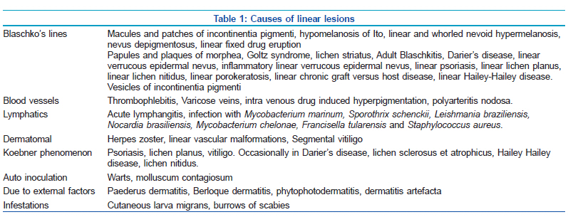
Blaschko’s lines are imaginary skin lines which various skin conditions are known to follow. They do not follow neural, vascular, or lymphatic structures and are distinct from dermatomes as well as Langer’s lines.[1] These were first described by Alfred Blaschko in 1901. The most widely accepted basis for these lines is genotypic mosaicism, i.e., presence of more than one type of cell lines within the body.[2,3] The causes for mosaicism are half chromatid mutation, lyonization, post zygotic mutation, chromosomal non disjunction, and chimerism.[3] The most common disorders following Blaschko’s lines are enlisted in Table 1.[2-5]
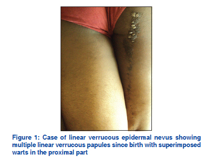
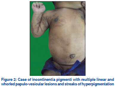
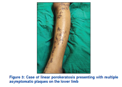
Figure 1, Figure 2, Figure 3 show a few conditions following Blaschko’s lines.
There are case reports of hypermelanotic macules of McCune-Albright syndrome,[6] extragenital lichen sclerosis,[7] eosinophilic cellulitis,[8] erythematous rash of scarlet fever,[9] Bart syndrome,[10] linear scalp lupus profundus,[11] all following Blaschko’s lines. BLOOD VESSELS Blood vessels have a longitudinal course in the body. Hence, lesions which occur in blood vessels are likely to have a linear pattern. These include the following:
- Thrombophlebitis refers to blood vessel inflammation along with thrombus formation. Two types are recognised - superficial and deep. Superficial thrombophlebitis is visible and presents with erythema and tenderness in the skin along the distribution of the affected blood vessel.[12] Mondor’s disease is a type of superficial thrombophlebitis which affects the breast and sometimes the penis, which presents with a linear and tender cord- like thickening of the affected vein.[13,14]
- Varicose veins present with dilated and tortuous veins commonly occurring in the lower limbs due to insufficient closure of valves causing backflow from deep to superficial veins.[15] Veins being linearly arranged, these lesions show a linear distribution.
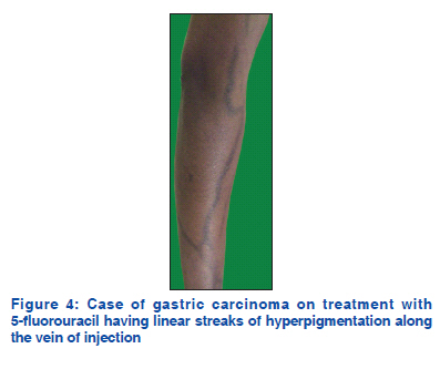
- Intravenous drug induced hyperpigmentation presents with linear hyperpigmented streaks along the course of veins. It commonly occurs in case of intravenous drugs given for malignancies[16] [Figure 4].
- Polyarteritis nodosa is a necrotising inflammation of blood vessels which presents with nodules and ulcers mainly on the lower limbs along the distribution of blood vessels.
LYMPHATICS
The following conditions follow lymphatics:
- Lymphangitis is the inflammation of lymph vessels. Acute lymphangitis is streptococcal infection of lymphatics of subcutaneous tissue presenting with erythematous linear streaks from the site of infection to the draining lymph nodes.[17] It also occurs in early stages of filariasis.
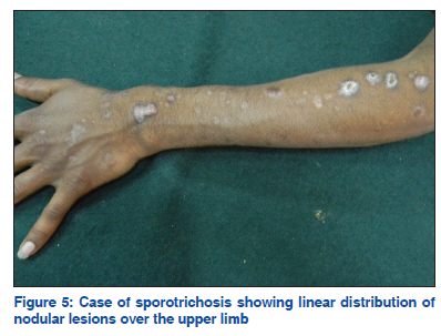
- Nodular lymphangitis is a clinical presentation in which erythematous papules and nodules occur along the course of lymphatics.[18] This occurs mainly in chronic suppurative inflammatory disorders of the skin in which causative organisms spread via lymphatics. This distribution commonly occurs in sporotrichosis and hence is also called sporotrichoid distribution [Figure 5]. The differential diagnosis for this includes infection with Mycobacterium marinum, Sporothrix schenckii, Leishmania braziliensis, Nocardia species, Mycobacterium chelonae, Francisella tularensis, and Staphylococcus aureus[18-21]
- DERMATOMES
A dermatome is a linear area of skin supplied by a single spinal nerve. The following conditions follow dermatomes:
- Herpes zoster presents with multiple erythematous
vesicles with burning sensation along the
distribution of a particular spinal nerve.
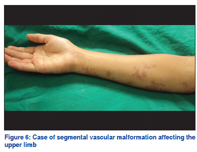
- Vascular malformations like capillary malformations
are sometimes are distributed along a dermatomal
segment[22] [Figure 6].
- Vitiligo is an acquired disorder which presents with depigmented macules and patches. Sometimes, this disorder follows dermatomal segments when it presents with linear macules and patches and is called as dermatomal, segmental, zosteriform or pseudosegmentalis type of vitiligo.[23] Segmental vitiligo has an early onset, rapid progression and no specific precipitating factors.[24] In one study, the most commonly involved dermatome was the trigeminal and only a few patients had an associated autoimmune disease.[24]
KOEBNER’S PHENOMENON
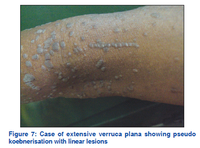
Koebner’s phenomenon was first described by Heinrich Koebner,[25] and refers to the development of isomorphic lesions at the sites of trauma in case of cutaneous disorders.[26] Scratching being a common cause of cutaneous trauma, leads to linear lesions along the line of scratching. True Koebner’s phenomenon occurs in psoriasis, lichen planus and vitiligo and is also occasionally seen in other conditions[26] [Table 1]. AUTO INOCULATION This is also termed as pseudo Koebner’s phenomenon. It occurs in infectious disorders like due to implantation of infectious agent in the skin during trauma leading to development of isomorphic linear lesions like in Koebner’s phenomenon. Examples include warts and molluscum contagiosum[26] [Figure 7].
EXTERNAL FACTORS
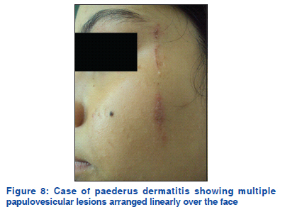
- Paederus dermatitis (dermatitis linearis,[27] or
blister beetle dermatitis[28]) commonly occurs in
tropical and subtropical regions. It is a peculiar
irritant contact dermatitis characterized by sudden
onset linear erythematous and bullous lesions and
burning on exposed areas of the body.[28] It occurs
when beetles of the genus Paederus (also called
rove beetles) are crushed on the skin, releasing the
vesicant pederin, which causes irritant dermatitis[29]
[Figure 8].
- Berloque dermatitis occurs when perfumes
containing bergamot oil are applied followed by
exposure to sunlight. It presents with erythema and
hyperpigmentation along the pattern formed by the
trickle of droplets, which is usually linear.[30]
- Phytophotodermatits (meadow dermatitis,
strimmer dermatitis, weed wacker dermatitis)[30]
occurs due to furocoumarins in plants which cause
a phototoxic reaction. It commonly presents with linear streaks along the area of contact with the
irritant.[31]
- Dermatitits artefacta: Dermatitis artefacta (factitious
dermatitis) refers to self-inflicted skin injuries
made consciously to elicit sympathy, escape
responsibility, or collect disability insurance. The
lesions are common on areas which are easily
accessible and commonly arranged in a linear
pattern.[32]
INFESTATIONS
- Cutaneous larva migrans is a skin infestation clinically characterized by erythematous linear serpiginous lesions caused by nematode larvae.[33]
- The burrows made by the scabies mite in the skin are also commonly linear.
CONCLUSION The importance of linear lesions in dermatology cannot be over emphasized. Linear lesions act as diagnostic clues in many disorders. They also help in elucidating the pathogenesis as they give a clue to the pathway of spread of the disease. Koebner phenomenon indicates the presence of active disease and helps to decide the line of management. REFERENCES
- Jackson R. The lines of Blaschko: A review and reconsideration: Observations of the cause of certain unusual linear conditions of the skin. Br J Dermatol 1976;95:349-60.
- Tagra S, Talwar AK, Walia RS. Lines of Blaschko. Indian J Dermatol Venereol Leprol 2005;71:57-9.
- Moss C. Mosaicism and linear lesions. In: Bolognia JL, Jorizzo JL, Rapini RP, editors. Dermatology. 2nd ed. Spain: Mosby Elsevier; 2008. p. 841-5.
- Bolognia JL, Orlow SJ, Glick SA. Lines of Blaschko. J Am Acad Dermatol 1994;31:157-90.
- Moss C, Larkins S, Stacey M, Blight A, Farndon PA, Davison EV. Epidermal mosaicism and Blaschko’s lines. J Med Genet 1993;30:752-5.
- Rieger E, Kofler R, Borkenstein M, Schwingshandl J, Soyer HP, Kerl H. Melanotic macules following Blaschko’s lines in McCune-Albright syndrome. Br J Dermatol 1994;130:215-20.
- Choi SW, Yang JE, Park HJ, Kim CW. A case of extragenital lichen sclerosus following Blaschko’s lines. J Am Acad Dermatol 2000;43:903-4.
- Sommer S, Wilkinson SM, Merchant WJ. Eosinophilic cellulitis following the lines of Blaschko. Clin Exp Dermatol 1999;24: 449-51.
- Duran-McKinster C, Moises C, Rodriguez-Jurado R, Tamayo-Sanchez L, Orozco-Covarrubias L, Ruiz-Maldonado R. Streptococcal exanthem in a blaschkolinear pattern: Clinical evidence for genetic mosaicism in hypomelanosis of ito. Pediatr Dermatol 2002;19:423-5.
- Duran-McKinster C, Rivera-Franco A, Tamayo L, De La Luz Orozco-Covarrubias M, Ruiz-Maldonado R. Bart Syndrome: the congenital localized absence of skin may follow the lines of
Blaschko: Report of Six Cases. Pediatr Dermatol 2000;17:179–82.
- Nagai Y, Ishikawa O, Hattori T, Ogawa T. Linear lupus
erythematosus profundus on the scalp following the lines of
Blaschko. Eur J Dermatol 2003;13:294-6.
- Torpy JM, Burke AE, Glass RM. JAMA patient page:
Thrombophlebitis. JAMA 2006;296:468.
- Pugh CM, DeWitty RL. Mondor’s disease. J Natl Med Assoc
1996;88:359-63.
- Kumar B, Narang T, Radotra BD, Gupta S. Mondor’s disease of
penis: A forgotten disease. Sex Transm Infect 2005;81:480-2.
- Nullen H, Noppeney T. Diagnosis and treatment of varicose
veins, Part 1: Definition, epidemiology, etiology, classification,
clinical aspects, diagnostic and indications. Chirurq
2010;81:1035-44.
- Alley E, Green R, Schuchter L. Cutaneous toxicities of cancer
therapy. Curr Opin Oncol 2002;14:212-6.
- Singh G, Kaur V, Singh S. Bacterial infections. In: Valia RG,
Valia AR, editors. IADVL Textbook of Dermatology. 3rd ed.
Mumbai: Bhalani Publishing House; 2008. p. 232.
- Giordano CN, Kalb RE, Brass C, Lin L, Helm TN. Nodular
lymphangitis: Report of a case with presentation of a diagnostic
paradigm. Dermatol Online J 2010;16:1.
- Schneider P, Monsel G, Veziris N, Roujeau JC, Bricaire F,
Caumes E. Successful treatment of nodular lymphangitis due to
Mycobacterium chelonae in two immunosuppressed patients.
Dermatol Online J 2011;17:8.
- Kostman JR, DiNubile MJ. Nodular lymphangitis: A
distinctive but often unrecognized syndrome. Ann Intern Med
1993;118:883-8.
- Lieberman AA, Grossman ME, Bloomgarden D. Sporotrichoid
lymphangitis due to Staphylococcus aureus in a diabetic
patient. Clin Infect Dis 1995;21:433-4.
- Boon LM, Vikkula M. Vascular Malformations. In: Wolff K,
Goldsmith LA, Katz SI, Gilchrest BA, Paller AS, Leffell DJ,
editors. Fitzpatrick’s Dermatology in General Medicine. 7th ed.
New York: McGraw Hill Book; 2008. p. 1652.
- Dhar S, Dutta P, Malakar R. Pigmentary Disorders. In: Valia
RG, Valia AR, editors. IADVL Textbook of Dermatology. 3rd ed.
Mumbai: Bhalani Publishing House; 2008. p. 753.
- Hann SK, Lee HJ. Segmental vitiligo: Clinical findings in 208
patients. J Am Acad Dermatol 1996;35:671-4.
- Kuner N, Hartschuh W, Khan-Durani B. Heinrich Kobner
and the “isomorphic phenomenon: History and review of the
literature. Hautarzt 2003;54:274-8.
- Thappa DM. The isomorphic phenomenon of Koebner. Indian J
Dermatol Venereol Leprol 2004;70:187-9.
- Morsy TA, Arafa MA, Younis TA, Mahmoud IA. Studies on
Paederus alfierii Koch (Coleoptera:Staphylinidae) with special
reference to the medical importance. J Egypt Soc Parasitol
1996;26:337-51.
- Singh G, Yousuf Ali S. Paederus dermatitis. Indian J Dermatol
Venereol Leprol 2007;73:13-5.
- Zargari O, Kimyai-Asadi A, Fathalikhani F, Panahi M. Paederus
dermatitis in northern Iran: A report of 156 cases. Int J Dermatol
2003;42:608-12.
- Anstey AV. Disorders of skin colour. In: Burns T, Breathnach S,
Cox N, Griffiths C, editors. Rook’s Textbook of Dermatology. 8th
ed. Oxford: Wiley Blackwell; 2010. p. 58.32.
- Adams SP. Dermacase: Phytophotodermatitis. Can Fam
Physician 1998;44:503,509.
- James WD, Berger TG, Elston DM, editors. Pruritus and
neurocutaneous dermatoses. In: Andrews’ Diseases of the Skin
Clinical Dermatology. 10th ed. Philadelphia: Saunders Elsevier;
2006. p. 61.
- Tomovic M, Skiljevic D, Zivanovic D, Tanasilovic S, Vesic S,
Dakovic Z, et al. Two cases of probable endogenous extensive
cutaneous larva migrans in Serbia. Acta Dermatovenerol Alp
Panonica Adriat 2008;17:37-40.
Copyright 2011 - Indian Journal of Dermatology, Venereology, and Leprology
The following images related to this document are available:
Photo images
[dv11219f7.jpg]
[dv11219f3.jpg]
[dv11219t1.jpg]
[dv11219f8.jpg]
[dv11219f2.jpg]
[dv11219f5.jpg]
[dv11219f1.jpg]
[dv11219f4.jpg]
[dv11219f6.jpg]
|
