 |
| About Bioline | All Journals | Testimonials | Membership | News |
|
||||||
|
||||||
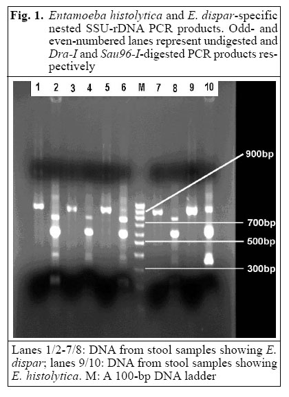
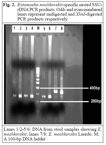
Journal of Health, Population and Nutrition, Vol. 23, No. 3, Sept, 2005, pp. 292-295 Short Report Entamoeba moshkovskii and Entamoeba dispar -associated Infections in Pondicherry, India Subhash Chandra Parija; Krishna Khairnar; Department of Microbiology, Jawaharlal Institute of Postgraduate
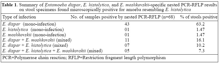
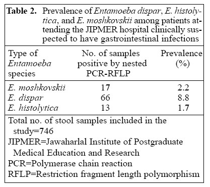
Medical Education and Research, Pondicherry 605 006, India Code Number: hn05037 ABSTRACT The prevalence of Laredo strain - Entamoeba moshkovskii - and non-pathogenic E. dispar in patients attending the Jawaharlal Institute of Postgraduate Medical Education and Research hospital, Pondicherry, India, is reported here. E. moshkovskii is reported for the first time in India. The species are morpho-logically indistinguishable from pathogenic E. histolytica. Of 746 stool samples screened, 68 showing cyst or trophozoite stage of E. histolytica, E. dispar, or E. moshkovskii were subjected to small sub-unit (SSU) rRNA gene-based polymerase chain reaction, which revealed a higher prevalence of E. dispar (8.8%) and E. moshkovskii (2.2%) compared to E. histolytica (1.7%) in patients. Only 19% of the 68 stool samples, resembling E. histolytica by microscopy, were actually E. histolytica , implying that 81% of suspected infections were misdiagnosed and would have been treated unnecessarily with antiamoebic drugs. Key words: Entamoeba histolytica; Entamoeba moshkovskii; Entamoeba dispar ; Amoebiasis; Diagnosis, Laboratory; India Introduction Entamoeba moshkovskii is primarily a free-living amoeba. It is indistinguishable in its cyst and trophozoite forms from E. histolytica, the causative agent of amoebiasis. E. moshkovskii has so far rarely been shown to infect humans (1). E. moshkovskii in humans has been reported from North America, Italy, South Africa, and Bangladesh, but it has never been associated with disease (2). The prevalence of E. moshkovskii in India has not been reported earlier. The morphological similarity of E. moshkovskii and E. dispar to disease-causing E. histolytica makes it important to differentiate the three species by polymerase chain reaction (PCR). In the clinical setting, this may lead to a misdiagnosis and unnecessary treatment with anti-amoebic chemotherapy (3). Materials and Methods Stool specimens for this study were obtained from patients attending the Jawaharlal Institute of Postgraduate Medical Education and Research (JIPMER) hospital, Pondicherry, India. In total, 746 stool samples from patients clinically suspected to have gastrointestinal infections were collected during July-December 2004 and were screened by microscopy, of which 68 showing tropho-zoite/cyst were subjected to E. histolytica, E. dispar, and E. moshkovskii-specific nested PCR. The DNA was isolated using the cetyltrimethylam-monium bromide (CTAB) extraction method (4). The extracted DNA was passed through DNA clean-up spin columns (Bangalore Genei, Bangalore) to remove PCR inhibitors. Based on the sequences of the small sub-unit (SSU)-rDNA of E. histolytica and E. dispar, nested sets of primers (designated E-1/E-2, Eh-1/Eh-2, and Ed-1/Ed-2) were used for detecting E. histolytica and E. dispar in stool specimens. The PCR was given a hot start by preincubating the PCR mix at 96 °C for two minutes, followed by 30 cycles - each consisting of 92 °C for 60s, 43 °C for 60s, and 72 °C for 90s and 72 °C for five minutes - one cycle for the final extension (5). In the nested PCR, annealing temperature was raised to 62 °C, leaving the other parameters of the amplification cycles unchanged. E. histolytica and E. dispar-specific nested SSU-rDNA gene amplification products were double-digested with restriction endonuclease Dra-I and Sau96-I for two hours at 37 °C according to the instructions of the manufacturer (Bangalore Genei) to verify the identity of species. Products were visualized on a 1.3% agarose gel containing ethidium bromide (0.2 mg/mL). The product of nested PCR from both E. histolytica and E. dispar showed 900-bp fragments which were further confirmed by restriction fragment length polymorphism (RFLP). The RFLP pattern for E. histolytica showed 550-bp and 350-bp fragments and undigested 900 bp, whereas for E. dispar it showed 700 bp, 550 bp, and confluent bands of 200 bp and 150 bp (Fig. 1). Based on the sequence of the SSU-rDNA gene of E. moshkovskii Laredo (GenBank accession no. AF 149906), a nested set of primers (designated Em-1/Em-2 and nEm-1/nEm-2) was used (6) for detecting E. moshkovskii in stool DNA. The PCR conditions were same as described above, except that the annealing temperature was 55 °C for the first PCR and 62 °C for the nested PCR. E. moshkovskii-specific nested SSU-rDNA gene amplification products were digested with restriction endonuclease XhoI for one hour at 37 °C according to the instructions of the manufacturer (Promega) to verify the identity of species. Products were visualized on a 1.8% agarose gel contain-ing ethidium bromide (0.2 mg/mL). The product of nested PCR from E. moshkovskii showed a 258-bp fragment which was further confirmed by RFLP. XhoI exclusively cuts the 258-bp product to produce 236-bp and 22-bp fragments (The 22-bp product is not visible in gel because it was too small to be resolved in 1.8% agarose gel). The standard strain of E. moshkovskii Laredo showed a higher molecular size (300 bp approximately) product compared to clinical isolates (Fig. 2). DNA from standard cultures of E. histolytica HM-1: IMSS, E. dispar SAW760, and E. moshkovskii Laredo was used as positive control, and stool samples showing no trophozoite/cyst were used as negative control. ResultsThe primer sequence for E. moshkovskii, E. histolytica, and E. dispar, blasted in the genome database of all organisms in the website (http://www.ncbi.nlm.nih.gov/blast/), was found to be specific for the study. Moreover, the amplified products of nested PCR were restriction-digested to rule out any non-specific amplification. The reference strain - E. moshkovskii Laredo - gave a band at approximately 300 bp with the E. moshkovskii-specific SSU-rDNA-nested primers, whereas the control - E. histolytica HM-1: IMSS and E. dispar SAW760 DNAs - was negative. The results of nested PCR-RFLP on the 68 stool DNA samples showing trophozoite/cyst resembling E. histolytica are shown in Table 1. One sample, negative by stool PCR for both E. histolytica and E. dispar, was eventually positive for E. moshkovskii. Sixteen of 17 E. moshkovskii-positive stool samples were also positive for E. histolytica, E. dispar, or both by SSU-rDNA PCR. Comparison of SSU-rDNA sequences from E. moshkovskii, E. histolytica, and E. dispar showed that the restriction endonuclease XhoI cut exclusively in the E. moshkovskii-specific, 258-bp - nested PCR product to pro-duce 236-bp and 22-bp fragments. Products from all the 17 positive stool samples and the Laredo strain showed the presence of this site (Fig. 2). The prevalence of E. moshkovskii, E. dispar, and E. histolytica among patients clinically suspected to have gastrointestinal infections, attending the JIPMER hospital, is shown in Table 2. Discussion The study was conducted to identify the prevalence of E. moshkovskii, E. dispar, and E. histolytica in stool samples of patients attending the JIPMER hospital. The study, for the first time, reports the prevalence of E. moshkovskii in India. The morphological similarity leads to confusion in diagnosis of amoebiasis in clinical settings. We have used nested PCR to detect infections due to E. histolytica, E. dispar, and E. moshkovskii because nested PCR increases the specificity and is more efficient in amplifying stool DNA. Our study included patients from varied age-groups and from different geographical localities, which shows the wide distribution of E. moshkovskii and E. dispar in Pondicherry and its neighbouring areas. The study has several interesting findings. Only one patient with dysentery showed E. moshkovskii-associated mono-infection. The cause remained undetermined; bacterial aetiology by routine stool culture was negative. Other investigations, such as viral study, could not be done in the laboratory. The high prevalence of E. moshkovskii among the study population supports the view that humans are a true host for this freeliving amoeba and are not just transiently infected (6). The study also answers the mystery of some microscopically-positive but antigenically- and PCR-negative results for E. histolytica and E. dispar (7), which, in our study, was due to E. moshkovskii. Eleven patients with mixed infections due to E. dispar and E. moshkovskii and five patients due to E. dispar, E. histolytica, and E. moshkovskii had no diarrhoea or dysentery, but had complaints of mild gastrointestinal discomfort. The study has shown appreciably a high prevalence of E. dispar and E. moshkovskii in the patients compared to E. histolytica which reveals that only 19% of the 68 stool samples, resembling E. histolytica by microscopy, were actually E. histolytica, implying that 81% of suspected infections were misdiagnosed and would have been treated unnecessarily with anti-amoebic drugs when diagnosed based on microscopic findings alone. Thus, epidemiologic studies and clinical diagnosis of E. histolytica-associated infection, which are based on morphological examination alone, are prone to error. Infections due to both E. dispar and E. moshkovskii are associated with asymptomatic carrier stage. The trophozoites of both E. dispar and E. moshkovskii lack the capability to invade the intestinal mucosa and do not have any ingested erythrocytes unlike that of E. histolytica. PCR is, therefore, essential to distinguish E. histolytica from E. dispar and E. moshkovskii. AcknowledgementsWe sincerely thank Dr. C. Graham Clark from London School of Hygiene & Tropical Medicine for providing us with lyophilized DNA of standard cultures of E. histolytica HM-1: IMSS, E. dispar SAW760, and E. moshkovskii Laredo References
© 2005 ICDDR,B: Centre for Health and Population Research The following images related to this document are available:Photo images[hn05037t2.jpg] [hn05037t1.jpg] [hn05037f1.jpg] [hn05037f2.jpg] |
| |||||||||
