 |
| About Bioline | All Journals | Testimonials | Membership | News |
|
||||||
|
||||||
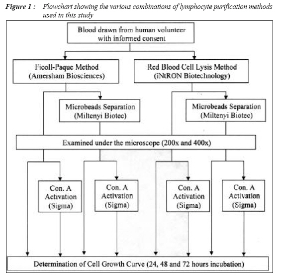
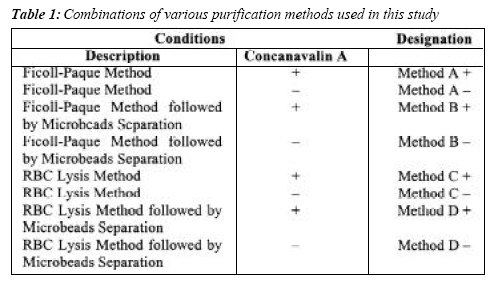
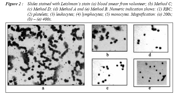
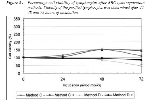
Malaysian Journal of Medical Sciences, Vol. 14, No. 1, Jan 2007, pp. 38-45 ORIGINAL ARTICLE DEVELOPMENT OF A PURIFICATION METHOD OF PURE PRIMARY LYMPHOCYTES FOR CELL VIABILITY ASSAYS Chan Kok Keong, Vishna Devi V Nadarajah* and Tay Ju Lee** Department of Postgraduate Studies and Research, *Department of Human Biology & Cells and Molecules, International Medical University, Sesama Centre, Plaza Komanwel, Bukit Jalil, 57000 Kuala Lumpur, Malaysia. Corresponding Author : Dr. Vishna Dewi V Nadarajah BSc(Hons(Mal.), Phd(Cantab), Department of Human Biology & Cells and Molecules, International Medical University, Sesama Centre, Plaza Komanwel, Bukit Jalil, 57000 Kuala Lumpur, Malaysia. Tel: 03-86567228 Fax: 03-86567229 E-mail: vishnadevi_nadarajah@imu.edu.my Submitted-20-02-2004, Accepted-03-12-06 Code Number: mj07007 The maintenance of pure primary lymphocytes culture for long periods may be difficult because of its inability to divide continuously. In addition, lymphocytes separation methods such as Ficoll-Paque, RBC lysis and immunomagnetic microbeads separation may have some affect on cell viability. The objective of this study is to determine various types of lymphocytes purification methods, in order to prolong primary lymphocytes culture to 72 hours. The second objective is to use these primary lymphocytes as targets for quantitative and qualitative cell viability assays when analysing the action of toxins isolated from natural products. Human blood was drawn and purified by using Ficoll-Paque, RBC lysis or immunomagnetic separation column method in various combinations. The purified lymphocytes were also grown with and without the growth enhancement factor, concanavalin-A. Cell viability assays were carried out for 72 hours at 24 hours interval. The lymphocytes purified using RBC lysis method, with or without concanavalin-A can prolong 100% cell viability for 72 hours whilst lymphocytes purified using Ficoll-Paque and supplemented with concanavalin-A showed an increase in cell viability of over 250% at 72 hours incubation. It was observed only lymphocytes purified using Ficoll-Paque followed by the immunomagnetic microbeads separation method and supplemented with concanavalin-A showed overall cell viability increase, reaching 300% at 72 hours incubation. This method was a reliable model to test the cytotoxicity of the Bacillus thuringiensis parasporal inclusion, suggesting that the method achieves the objectives of the study. Keywords: lymphocytes, purification, cell viability Introduction In medical research, the cellular component of blood is one of prime interests, for example, to determine the efficacy and cytotoxicity of natural products. Yet, most of the blood sample separation procedures involve methodologies prone to introducing artifacts, and require skilled technicians as well as equipped and expensive laboratories (1). Several protocols and products on lymphocytes purification have been developed, such as Ficoll- Paque (2, 3), Red Blood Cell (RBC) lysis (4) and immunomagnetic microbeads separation (5,6). These protocols have been used either alone or in combination to obtain pure lymphocytes (2, 4–7). However, the major disadvantage of these protocols is poor recovery of the target lymphocytes resulting in low yields. To overcome the low yield, the lymphocytes are cultured as cell proliferation will result in a higher yield. Nevertheless, the maintenance of pure primary lymphocytes culture for long periods may be difficult because of its inability to divide continuously in vitro (8,9). Moreover, the above mentioned purification protocols may have some impact on the lymphocytes microenvironment during separation and that may affect the cell viability. Due to this reason, it is vital to improve the turnover of lymphocytes after separation in order to enhance the life span of lymphocytes. The present study aims to compare the various types of lymphocytes purification methods, in order to find a suitable protocol that can prolong primary lymphocytes culture to 72 hours. This is important if the purified primary lymphocytes are to be used as targets for quantitative and qualitative cell viability assays for the analysis of toxins isolated from natural products. Methodology Lymphocytes Purification and Activation Whole blood was taken from healthy human volunteers with informed consent. Four combinations of lymphocyte purification procedures are conducted in parallel experiments (as shown in Figure 1) to determine the purification efficiency. Purification efficiency was determined by compound microscope (Leica DMLS, Leica Microsystems, Wetzlar GmbH, Germany). Table 1 summarizes the four purification combinations into Methods A, B , C and D. Each of the four combinations were further divided into two sub-groups, whereby concanavalin A (Con. A) was added to one of the sub-groups to activate the lymphocytes. Purification protocols provided by the manufacturer were adapted with slight modification to suite the local environment, i.e. the purification process was done in 4oC rather than room temperature. The purity of lymphocytes was determined using microscope. Determination of Cell Growth CurveThe purified lymphocytes which were separated separated using the combinations used in Table 1, were counted using haemocytometer (Hawksley& Sons Ltd., England, United Kingdom) and diluted to the appropriate concentration to achieve a seeding density of 1 x 10⁶ viable cells/ml. 100_l of the thoroughly mixed suspension was seeded in triplicates into the wells of the 96-wells plate and the plates were then incubated. Con. A (100mg/ml) was added to the sub-groups in order to activate the lymphocytes. Plates were taken out after 24, 48 and 72 hours of incubation and 3-(4,5-dimethylthiazol- 2-yl)-2,5-diphenyl tetrazolium bromide (MTT) cell viability assay was carried out as described by Mosmann (10) with slight modifications, i.e. hydrochloric acid isopropanol has been added into the wells, whereby the hydrochloric acid convert the phenol red indicator in tissue culture medium to a yellow color that does not interfere with MTT formazan measurement while the isopropanol dissolves the formazan to give a homogeneous blue solution suitable for absorbance measurement. The absorbance were then measured using ELISA plate reader (Expert Plus UV, Asys Hitech GmbH, Austria) with a test wavelength of 570 nm and a reference wavelength of 630 nm (Figure 1). Solubilisation and Activation of Bacillus thuringiensis toxinsToxins harvested from sporulated Bacillus thuringiensis were solubilized with 100 ml sodium carbonate and 100 ml DTT followed by incubation at 37oC water bath for one hour. After incubation, the solubilised toxins were activated by adding 120 ml trypsin and incubated again at 37oC water bath for one hour. The solubilised and activated toxins were collected by centrifugation at 13,000 rpm for two minutes, followed by filtration with 0.2 mm filters. Cell Viability AssayIn the cell viability assay, the purified lymphocytes which gave the best growth curve were selected to be treated with cytotoxic and non-cytotoxic natural products to determine the usability of the purification method on cell viability assay. The natural products used in this study were proteins isolated from Malaysian strains of Bacillus thuringiensis, which are currently being studied for its in vitro effects on leukaemic T cells (11–13). The purified lymphocytes were counted and diluted to the appropriate concentration to achieve a seeding density of 2 x 10⁶ viable cells/mL. 50_l of the wellmixed suspension was seeded in triplicates into the wells of the 96-wells plate and incubated for one hour in order to let the cell settle. The treatment solutions were made up to 1024 mg/ml in cell culture medium. The stock treatment solutions were then adjusted to 1024, 512, 256, 128, 64, 32, 16, 8, 4, 2 and 1 mg/ml, using serial dilution method to obtain the above treatment concentration. 50µl of each treatment solution was then dispensed into each of the wells in triplicates. The final volume in each well was 100 ml and the treatment concentration was 512, 256, 128, 64, 32, 16, 8, 4, 2, 1 and 0.5 mg/ml. A plate was taken out after 24, 48 and 72 hours of incubation and MTT cell viability assay was carried out. Statistical AnalysisStatistically significant differences were evaluated by one way analysis of variance (ANOVA). Statistical analysis was performed using SPSS statistical software (version 13, SPSS Inc., Chicago, Illinois, USA). Results and DiscussionLymphocytes were separated from the blood of normal volunteers for use in Method A, B, C and D. When compared to a blood smear shown in Figure 2a, all methods show ability to isolate lymphocytes (Figure 2b & c). However, as shown in Figure 2b, the Method C shows the presence of monocytes and cell debris. Cell debris maybe generated when the RBC lyses and sediments together with the lymphocytes during centrifugation. On the other hand, Method A shows a ‘cleaner’ preparation as cell debris were not generated, however, some platelets were found in the preparation as mentioned by Boyum (3). The immunomagnetic microbeads separation methods gave satisfactory results in either Method D (Figure 2d) or Method B (Figure 2e). This negative sorting method (6) filtered out the cell debris and platelets, but it trapped the monocytes using magnetic conjugated specific antibodies. In cell growth assay, percentage cell viability of lymphocytes decreased significantly (p < 0.05) for Method A –, B – and D – at 24, 48 and 72-hours incubation except for Method C – (Figure 3 and 4). This could be due to the growth of other mononuclear cells that does not require supplements. Method A +, B +, C + and D + generally showed significant (p < 0.05) increase in percentage of cell viability compared with Method A –, B –, C – and D –. These results confirm that concanavalin A is a mitogen that stimulates growth of human lymphocytes, which in agreement to the findings, using chicken as a model as reported by Toivanen and Toivanen (14). The results also agree with earlier findings that concanavalin A stimulates lymphocytes to undergo blastogenic transformation and synthesize of DNA, in vitro (15). The supplemented lymphocytes cultures in Method A or Method B showed significantly (p < 0.05) better growth than lymphocytes in Method C and Method D. Lymphocytes for the latter methods showed better growth on 24-hour incubation but the cell viability dropped significantly (p < 0.05) on 72hour incubation. These results shows that the RBC lysis method may have some impact on the cell microenvironment during the separation process and that may affect the cell viability, with or without supplement. Nevertheless, results show that lymphocyte cultures in Method B + display constant growth within the 72-hour incubation period. Hence it is suggested that this method is optimum and reliable for testing the effects of any potential therapeutic agent on normal T-lymphocytes. The results of cell viability assay which determines the effect of cytotoxic (Bt 4) and non-cytotoxic (Bt 18) (13) on normal lymphocyte culture in Method B + is shown in Figure 5, 6, 7. Treatment of purified and supplemented lymphocytes with various doses of Bt 4 (0–512mg/ml) following a 24-hour incubation period showed that Bt 4 significantly (p < 0.05) decreased the percentage cell viability of said lymphocytes from 85.71 to 60.71%. The above pattern of decrease in percentage of cell viability of purified and supplemented lymphocytes treated with Bt 4, are similar following 48- (Figure 6) and 72-hour (Figure 7) incubation periods. The cell viability of purified and supplemented lymphocytes treated with Bt 4, following 48-hour incubation period decreased the percentage cell viability of said lymphocytes from 76.7 to 23.3%. Similarly, the percentage cell viability decreased from 75.2 to 4.0% for purified and supplemented lymphocytes treated with Bt 4 following 72-hour incubation periods. This indicates that using lymphocytes isolated by method B +, there is a constant trend of Bt 4 action even though it is assayed at 24, 48 or 72 hours. On the other hand, treatment of normal lymphocyte culture Method B + with various doses of Bt 18 (0 – 512 mg/mL) following 24- to 72-hour incubation periods did not show any significant (p > 0.05) decrease in cell viability. This indicates that the lymphocytes prepared using method B + can be used reliably to distinguish between a cytotoxic toxin (Bt 4) and a non-cytotoxic inclusion protein (Bt 18). Thus, such treatment can produce lymphocytes which can be used as targets for quantitative and qualitative cell viability assays when analyzing the action of toxins isolated from natural products. Overall, method B gives lymphocytes of satisfactory purity for culture compared to the other methods. This separation method followed by enrichment with concanavalin A (mrethod B +), produced a culture with increasing cell viability to 300% of the initial counts within the 72-hour incubation period. Hence it is suggested that method B + is suitable to be used as targets for quantitative and qualitative cell viability assays. AcknowledgementThis study was a pilot study of project titled “The effects of Bacillus thuringiensis 18 parasporal inclusions on leukaemic cell viability and apoptosis, in vitro” supported by Grant No: IMU 082/2005 from the International Medical University, Bukit Jalil, Kuala Lumpur, Malaysia. Reference
© Copyright 2007 - Malaysian Journal of Medical Science The following images related to this document are available:Photo images[mj07007t1.jpg] [mj07007f3.jpg] [mj07007f4.jpg] [mj07007f7.jpg] [mj07007f5.jpg] [mj07007f2.jpg] [mj07007f1.jpg] [mj07007f6.jpg] |
| |||||||||