 |
| About Bioline | All Journals | Testimonials | Membership | News |
|
||||||
|
||||||
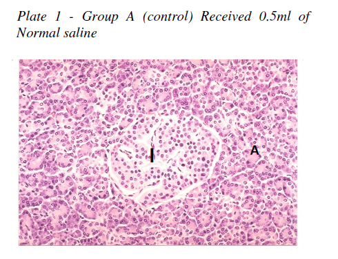
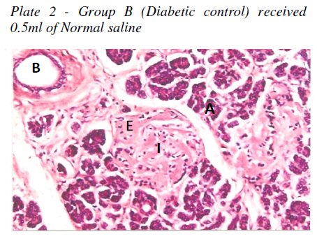
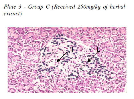
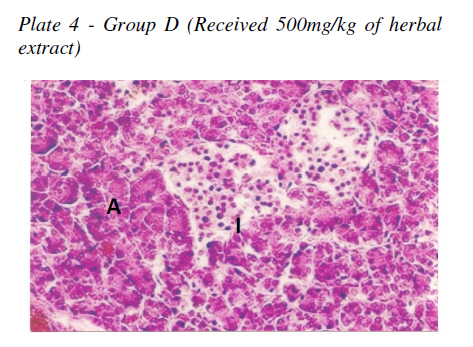
Nigerian Journal of Physiological Sciences, Vol. 24, No. 2, July-December, 2009, pp. 153-155 Original Article Histological changes in the pancreas following administration of ethanolic extract of Alchornea cordifolia leaf in alloxan-induced diabetic Wistar rats CF Eliakim-Ikechukwu, AI Obri Department of Anatomy, Faculty of Basic Medical Sciences, University of Calabar, Calabar, Nigeria Correspondence Address:C F Eliakim-Ikechukwu, Department of Anatomy, Faculty of Basic Medical Sciences, University of Calabar, Calabar, Nigeria, chisom_favor@yahoo.com Date of Submission: 20-Nov-2009 Code Number: np09026 Abstract Twenty-four wistar rats weighing between 150 and 170g were used in this research work. They were divided into four groups A, B, C, and D of six rats each. Diabetes was induced in Groups B, C, and D using a single intraperitoneal injection of 150mg/kg of Alloxan after an overnight fast. Group A served as the normal control while Group B served as the diabetic control and each received 0.5ml of normal saline daily. Groups C and D received 250mg/kgbwt/day and 500mg/kg /day of the plant extract respectively through orogastric intubation. All the animals were given normal rat chow and water freely. The experiment lasted for 28 days. The animals were anaesthetized using chloroform inhalation and the peritoneum stripped open and the pancreas removed and prepared for histological observation using haematoxylin and eosin staining technique. Histology showed regenerative changes of pancreatic islet cell at a dose of 500 mg/kg/day. Keywords: Pancreas, Alchornea cordifolia, diabetes mellitus, Wistar rats Introduction Alchornea cordifolia belongs to the family Euphorbiaceae. In Cross River State of Nigeria it is called ′Mbom′ by the Efiks and ′Ashenshen′ by the Bekwarra people. It has been used widely in the treatment of rheumatism, arthritis, colds, muscle pains, cough, infertility, impotence, diabetes and diarrhoea (Taylor, 2005). Phytochemical screening revealed the presence of alkaloids, saponins, tannins (naturally occurring polyphenol), flavonoids, terpenes and glycosides with steroidal rings (Olaleye et al, 2007) and Osadebe and Okoye, (2003). It is reasonably safe with an LD50 of the methanolic extract 1131.4mg/kg. Among its activities is its anti-inflammatory action which is only attributed to the terpenoid content (Osadebe and Okoye, 2003). It is one of the herbs found useful in traditional medicine practice in the management of diabetes but the scientific basis for this action has not yet been explored. Diabetes mellitus is a chronic metabolic disorder suffered by a high percentage of the world population. WHO estimates a likely increase in prevalence by 35% from about 171 million to 300million people in the year 2030. In the laboratory, experimental diabetes is commonly induced by using Alloxan or streptozotocin which selectively destroy the pancreatic B cells via production of reactive oxygen species. Alloxan induces type 1 DM while streptozotocin induces both type 1 and type 2 diabetes mellitus (Szkudelski, 2001). Insulin, whose absolute or relative lack leads to diabetes is produced by the B cells of pancreatic islets of Langerhans. The islets which represent the endocrine part of the pancreas contain two main cell types, the alpha (A) cells and the beta (B) cells. A third less common type is the delta (D) cell, and a fourth, very rare cell is the C-cell. The A cells which produce glucagon make up about 20% of the islet cells and have a characteristics peripheral distribution within the islet. The B cells which produce insulin are numerous forming about 70% of the islet cells and occupy the interior of the islet (Rhodin, 1974 and Wheater et al, 1987). This study will aim at validating or rejecting the claims of A. cordifolia in the management of diabetes mellitus. Materials and Methods Alchornea cordifolia leaves were picked from a garden in Calabar, Nigeria and taken to the University of Calabar botanical garden for identification. The leaves were washed free of debris and dust particles and were air dried at room temperature for two weeks. The dried leaves were blended into dry powder. 1000g of the powder were soaked in 2 litres of 98% ethanol for 72 hours and was thereafter filtered using Whatman No. 1 filter paper. The filtrate was concentrated using a rotary evaporator to yield 67g of extract (6.7% yield). This method was adopted from Ugochukwu and Babady, (2003). 20g of extract was dissolved with normal saline up to 100ml. Twenty-four wistar rats weighing between 150g to 170g were randomly divided into four groups: A, B, C and D. Group A served as the normal control and received 0.5ml of normal saline daily. Diabetes was induced in groups B, C and D using Alloxan, a uric acid derivative. The animals were fasted overnight and a single interperitoneal dose of 150g/kgbwt of Alloxan was used to induce each animal in groups B, C and D. After seven days of diabetes induction, fasting blood glucose (FBG) was done and only animals with FBG 20mmol/l and above were selected for the study. Group B served as the diabetic control and received only 0.5ml of normal saline daily. Groups C and D received daily doses of 250mg/kg and 500mg/kg of the herbal extract respectively via orogastric intubation. The administration lasted for 28 days. Thereafter the animals were anaesthetized using chloroform inhalation. The peritoneum was stripped open and the pancreas quickly harvested. The tissues were processed histologically and haematoxylin and eosin staining technique was employed, (Lillie, 1954). Result Plate 1 -Group A (control) Received 0.5ml of Normal saline A - Acinar cells, I - islet of Langerhans Cells of the pancreas were all present in their normal proportions. The acinar cells which stained strongly are arranged in lobules with prominent nuclei. The islet cells are seen embedded within the acinar cells and surrounded by a fine capsule. Magnification 100x. Plate 2 -Group B (Diabetic control) received 0.5ml of Normal saline E -Eosinophilic material, B - Blood vessel, Magnification 100x The acinar cells around the islets though seem to be in normal proportion does not look classical. The islets are largely occupied by a uniform eosinophilic material and few atrophic cells. Eosinophilic materials also surround the blood vessel. Plate 3 -Group C (Received 250mg/kg of herbal extract) The acinar cells were seen to be normal. The islets present with heavy lymphocytic infiltration in and around it (insulinitis). Some normal islet cells are also present. L - Lymphocyte, Magnification 100x Plate 4 -Group D (Received 500mg/kg of herbal extract) The acinar cells are seen to be normal. The islets are present with a large proportion of islet cells though with a smaller volume as compared with control. There is very scanty inflammatory cell infiltration and no eosinophilic deposits were seen. Magnification 100x. Discussion The majority of islet cells is formed by B cells which are responsible for producing insulin. Depletion of B cells will therefore result in insulin deficiency which will lead to a disorder in carbohydrate, protein and fat metabolism with a resultant hyperglycaemia. In this study, Alloxan which selectively destroy B cells of the islet was used to induce type 1 diabetes mellitus. Insulinitis and loss of B cells were observed which may be seen in type 1 DM. insulinitis is evidenced by heavy lymphocytic infiltration in and around the islet. This is commonly seen in islets containing residual B cells and it supports the possibility of a specific, immunologically mediated destruction of B cells as the cause of type 1 DM, (Anderson, 1986). There is no evidence of immune involvement in the pathogenesis of type 2 DM, (Kumar and Clark, 2005). Insulinitis was seen in the group treated with 250mg/kg/day of plant extract showing that some of the scanty cells seen in the islet are B cells. Large deposits of a homogenous eosinophilic material largely occupying the islet and around blood vessels are seen in the diabetic control group. This could be a localized amyloidosis which has been documented to occur in the pancreas in many diabetics. This is deposited extracellularly and first appears in the walls of small vessels. It is also deposited in reticulin fibres and basement lamina. When present in large amount, it induces pressure atrophy of the surrounding cells (Anderson, 1985). It is not surprising therefore that scanty atrophic cells are found in this group making worse the situation. This is related to chronic inflammatory disorder, (Kumar and Clark, 2005). Islet cells of group D treated with 500mg/kg/day of plant extract has regenerated considerably suggesting the presence of stable cells in the islets with the ability of regenerating (De Fronzo et al, 1997). This also suggests that the plant extract at this dose has the ability of inducing the quiescent cells to proliferate to replace the lost cells. The exact mechanism is not known but it has been documented that the flavonoid fraction of this plant extract decreases blood glucose and increases the number of B cells, (Chakrvarthy et al, 1980). It has also been documented that phenolic content of therapeutic plants contributes immensely to antioxidant activity of plants. The phenolic constituent of this plant may have stopped further destruction of the remaining B cells in the islet by mopping up the circulating reactive oxygen species generated by the alloxan to destroy the B cells and then allowing other phytochemicals of the plant to induce regenerative activities. Though the exact mechanism is unknown, it is obvious that A. cordifolia plant extract is able to cause regeneration of pancreatic B cells at a dose 500mg/kg/day either by its active constituents acting single or together. [13] References
Copyright 2009 - Nigerian Journal of Physiological Sciences The following images related to this document are available:Photo images[np09026f1.jpg] [np09026f2.jpg] [np09026f4.jpg] [np09026f3.jpg] |
| |||||||||
