 |
| About Bioline | All Journals | Testimonials | Membership | News |
|
||||||
|
||||||
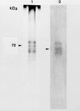
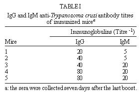
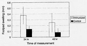
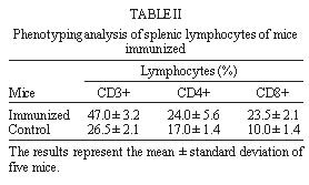
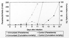
Mem Inst Oswaldo Cruz, Rio de Janeiro, Partial Protection of Mice against Trypanosoma cruzi after Immunizing with the TcY 72 Antigenic Preparation Yara M Gomes/+, Frederico GC Abath, Mineo Nakazawa, Paola Minoprio*, Ioannis Vouldoukis**, Loic Monjour** Departamento de Imunologia, Centro de Pesquisas Aggeu
Magalhães-Fiocruz, Av. Moraes Rego s/n, Cidade
Universitária, 50670-420 Recife, PE, Brasil + Corresponding author. Fax: +55-81-453.1911. E-mail: yara@cpqam.fiocruz.brReceived 9 February 1998 Code Number:OC99032 A 72 kDa Trypanosoma cruzi glycoprotein recognized by the 164C11 monoclonal antibody (IgM isotype) was purified by preparative electrophoresis. The antigenic preparation obtained, named TcY 72, was used to immunize C57Bl/10 mice. The following results were observed after immunization: (1) induction of higher titres of IgG than IgM antibodies, as evaluated by indirect immunofluorescence; (2) significant DTH after injection of epimastigotes in mice footpads; (3) peak parasitemia in immunized mice was significantly reduced and animals were negative by 13 days post-infection, although the mice still succumb to infection; (4) the phenotypic analysis of spleen cell populations showed a decrease in the CD4/CD8 ratio in immunized mice. Taken as a whole, these findings indicate that TcY 72 is immunogenic and potentially important for protective immunity. Key words: Trypanosoma cruzi - immunization - TcY antigenic preparation - Chagas' disease Chagas' disease, caused by the protozoan Trypanosoma cruzi, is distributed in South and Central America where 18-20 million people are affected (WHO 1991). No vaccine or safe chemotherapy is currently available for the prevention or treatment of this disease, which results in high morbidity of infected individuals. The use of insecticides to control the insect vector is a continuous source of concern because of the impact on the environment. New vaccination strategies for the better control of tropical diseases (Capron & Dessein 1988) and the recent knowledge of the complexity of parasite antigenic structure and of the immunopathology of the infectious process (Peterson et al. 1986, Choromanski & Kuhn 1987, Reed 1988) should renew research activity on vaccines against Chagas' disease. Many reports have demonstrated that partial protection of mice against T. cruzi may be induced by immunization with killed (Menezes 1968), attenuated (McHardy & Elphick 1978), or chemically inactivated (Andrews et al. 1985) parasites. More defined antigens such as purified glycoproteins isolated from the epimastigote membrane (Snary 1983), flagellar fraction (Segura et al. 1977, Wrightsman et al. 1995) and exoantigens (Gruppi et al. 1994, 1995) have been shown to have protective potential. The immune mechanisms responsible for resistance are still unclear. However, resistance to infection has been correlated with a decrease in polyclonal lymphocyte responses induced by the parasite (Minoprio et al. 1988). Recently, we obtained a monoclonal antibody (mAb) 164C11 which was generated from mice immunized with freeze-thawed and sonicated bloodstream trypomastigotes (Y strain) (Gomes et al. 1995). This mAb recognizes a single band with an apparent molecular weight of 72 kDa in Western blots obtained from all developmental stages of T. cruzi (Gomes et al. 1995). Studies using periodate and endoglycosidase treatments suggest that the epitope recognized by this mAb is not a carbohydrate and seems to be located on the parasite membrane. In addition, the 72 kDa protein seems be involved in adhesion/or internalization of bloodstream trypomastigotes in Vero and macrophage cells (Gomes et al. 1995). In the present report a fraction containing the 72 kDa protein (TcY 72) was isolated by preparative electrophoresis and its protective potential was evaluated in mice. MATERIALS AND METHODS Parasites - Epimastigote forms of T. cruzi Y strain, classified as Type I (Andrade 1985) were obtained from acellular cultures as previously described by Gomes et al. (1995), and bloodstream trypomastigotes were obtained from peripheral blood of Swiss-Webster mice acutely infected with the parasite. Purification of monoclonal antibody - 164C11 hybridoma cells secreting IgM (Gomes et al. 1995) were injected intraperitoneally into pristane-primed Balb/c mice. Ascitic fluid was collected 10-14 days later and clarified by centrifugation. The mAb was purified by gel filtration according to Bouvet al. (1984). The fraction containing IgM was analyzed by SDS-PAGE and Western blot. Antigenic preparations - The antigenic preparation was obtained by preparative electrophoresis according to the method proposed by Monjour et al. (1988). T. cruzi Y strain epimastigotes were lysed in Laemmli buffer containing protease inihibitors (1 mM PMSF, 0.1 mM EDTA - Sigma Chemical Co., St. Louis, Mo.). The lysates were separated in 10% SDS-PAGE (Laemmli 1970). The 72 kDa band was removed, sliced and electroeluted according to Andrews (1981). The purity of the electroluted band was analyzed by electrophoresis and the gel was silver-stained by the method of Morrisey (1981). Western blot - TcY 72 as well as the purified mAb containing the protease inhibithors (1 mM PMSF, 0.1 mM EDTA) were solubilized with sample buffer. TcY 72 and the purified mAb were then blotted onto NTC (0.45 mm pore size, Bio-Rad, Richmond, Ca) and the immunoassay performed according to Towbin et al. (1979). The strips containing the 72 kDa protein were incubated with the purified mAb followed by incubation with horseradish peroxidase-labeled IgM (heavy chain specific). To assess the purity of the purified mAb, a direct immunoassay was carried out using several anti-isotypes (aIgM, aIgA, aIgG1, aIgG2a, aIgG2b, aIgG3 - Caltag). The strips were developed with 0.01% DAB, 0.01% H2O2 in 100 mM PBS pH 7.3. Mice immunization - Two groups (G1 and GII) of 10 inbred C57Bl/10 female mice (17 ± 2 g) were used. G1 was injected three times (20 days apart) with 25 mg of TcY 72 subcutaneously. The first injection was emulsified in complete Freund´s adjuvant and the following in incomplete Freund´s adjuvant. G2, control mice, were injected with the elution buffer and the adjuvants. Five mice of each group were used for evaluation of antibody and delayed hypersensitivity responses and challenge experiments. The remaining five mice were used for the phenotyping of splenic cells. Antibody response - The antibody response of immunized mice was analyzed by indirect immunofluorescence (IIF) on Y strain formalized epimastigotes (Camargo 1966) seven days after the last boost. Rabbit a-mouse IgG and IgM conjugated to fluorescein were obtained from Sigma. Delayed-type hypersensitivity test (DTH) - DTH test was assayed in each animal 10 days after the last boost of immunization. Epimastigotes in exponential growth were killed by incubation in 1% buffered formalin and washed five times by centrifugation in 0.15 M NaCl pH 7.2. Ten million parasites in 50 ml were injected into the right footpad of each mouse from both immunized and control groups and NaCl was injected into the contralateral footpad. Swelling in both footpads was measured at 24 and 48 hr with a caliper (Mitutoyo-Japan). The results were reported as the difference between the swelling of the footpad injected with antigen and the swelling of the footpad injected with diluent. Infection - Twenty five days after the last boost, the animals were challenged with an intraperitoneal inoculation of 103 bloodstream trypo-mastigotes (Y strain). Parasitemia was determined by using samples of 5 ml of tail blood as previously described (Brener & Chiari 1963). Mortality was recorded daily. Membrane phenotyping of splenic cells - For this study five C57Bl/10 female mice were used, as described above and 20 days after the last boost the spleen was removed and collected in a sterile disposable 60 mm Petri dish containing 5 ml of RPMI 1640 medium (Sigma Chemical Co, St. Louis, Mo). The spleen cells were prepared by gently teasing and isolated by Histopaque-1077 (Sigma Chemical Co, St. Louis, Mo) density gradient centrifugation. The cells at the ring were collected, washed and suspended in RPMI at a concentration of 106 cells/ml. The cellular suspension (15 ml) was loaded on a glass slide and fixed in acetone. The detection of CD3+ T lymphocytes was performed by direct immunofluorescence with an anti-CD3 mAb (145-2c II, Boehringer Mannheim Biochemicals, Indianapolis, In) conjugated to FITC and diluted 1:40 in PBS pH 7.3. After incubation at 37oC for 30 min with 20 ml of mAb, the cells were washed and observed with a fluorescence microscope. The detection of CD4+ and CD8+ T lymphocytes was performed by using the avidin-biotin peroxidase system (Kit ABC, Vectastain PK 4002 - Vector Labor, Burlingane) according to the instructions of the supplier using the mAbs anti-CD4 (YTS - 191.1, Cedarlane) or anti-CD8 (53 - 6.7, Boehringer Mannheim Biochemicals, Indianapolis, In). Statistical analysis - Statistical analysis was performed by using Student's t test (Logiciels: Statgraphics - Statistical Graphics Corporation and Harvard - Microsoft Corporation). RESULTS Antigenic preparation - The TcY 72 was analyzed by electrophoresis and Western blot. Three polypeptides of 64, 72 and 82 kDa appeared on the gel (Figure 1). This result was unexpected and led us to propose some possible explanations: the 64 and 82 kDa polypeptide may be produced by partial degradation of protein followed by aggregation of the peptide fragments. Some membrane proteins are known to aggregate even in the presence of detergents including SDS (Helenius & Simons 1975, Dohnal et al. 1980). The fact that the purified mAb recognizes the polypeptides by Western blot (Fig. 1) supports our explanation. In this assay no reactivity was observed when normal mouse serum was used. However, as the possibility of copurification of other proteins comigrating with the 72 kDa glycoprotein was not excluded we will refer to the purified fraction as TcY 72 antigenic preparation. Antibody and cellular responses of immunized mice - The anti-T. cruzi antibody response as detected by IIF showed lower titres of IgM antibodies in comparison to IgG (p<0.025) (Table I). IgG and IgM anti-T. cruzi antibodies were not detected in the control. The delayed hypersensitivity reaction was strong when compared to the control. Significant 24 hr swelling (p<0.05) was present in all immunized mice slightly decreasing by 48 hr (Fig. 2). The phenotypic analysis of spleen cell populations at day 20 after the last boost revealed that CD3+ T cells (CD4+ and CD8+) increased in numbers when compared to normal mice (p<0.01). CD8+ T cell numbers were higher in the immunized group in comparison to the control (p< 0.01). Although CD4+ T cell numbers were also higher in the immunized group in comparison to the control, this difference was not shown to be statistically significant (p=0.17) (Table II). Parasitemia and cummulative mortality - The course of parasitemia in immunized mice is shown in Fig. 3. The parasitemia was lower on days 11 after infection (p<0.05) in comparison to non-immunized mice and complete clearance of parasites from the blood was observed by day 13 after challenge. The non-immunized mice developed high parasitemias and 100% mortality was observed day 11 (Fig. 3). While parasites were cleared by day 13 post-challenge, the immunized mice still succumb to infection by day 22. Nevertheless, death was significantly delayed in the immunized animals. FIGURE 1: electrophoresis and Western blot of the TcY72.(1) Electrophoretic profile after silver staining;(2) antigenic profile after the reaction with the purified mAb. No reaction was evidenced with the mouse normal serum (data not shown). The arrow shows the 72kDa band. TABLE I: IgG and IgM anti-Trypanozoma cruzi antibody titres of immunized micea FIGURE 2: delayed-type hypersensitivity response in mice immunized with TcY 72 antigen. Each bar represents the mean (± standard deviation) footpad swelling. TABLE II: Phenotyping analysis of splenic lymphocytes of mice immunized FIGURE 3: parasitemia and cumulative mortality of mice after challenge with 103 bloodstream trypomastigotes of Y strain. Each point represents the parasitemia mean of five mice. DISCUSSION In the present report, we analyzed the immunizing potential of the 72 kDa glycoprotein (identified by the 164C11 mAb raised against bloodstream Y strain trypomastigotes) that appears to be located on the parasite membrane and seem to be involved in adhesion or internalization of the parasite (Gomes et al. 1995). This glycoprotein was obtained by preparative electrophoresis and used to immunize C57Bl/10 female mice. Mice immunized with the TcY 72 antigenic preparation developed partial resistance to infection showing lower peak levels of parasitemia and longer survival times than non-immunized mice. These results are relevant if one takes into account that the Y strain used in the challenge is extremely virulent and leads to early death in naive animals (7-12 days post-infection). We are planning to repeat these experiments with a less pathogenic strain of the parasite. The antibody response evaluated by IIF revealed that immunized mouse sera contains IgM and IgG antibodies specific for the 72 kDa glycoprotein. In addition, a strong DTH reaction was observed suggesting that both responses play a role in protective immunity. It is possible that a 72 kDa glycoprotein identified by a mAb (WIC 29.26) produced against epimastigotes from a Y strain clone (Wel tryp Y2.CL) (Snary et al. 1981) shares similarities with the antigenic structure recognized by the mAb described here. However, previous results showed that this 72 kDa protein (Snary et al. 1981) failed to induce protection against a T. cruzi infection in terms of parasitemia and survival rates. Our results show partial but significant protection when the TcY 72 antigenic preparation is used in our mouse model. We show that mortality is delayed in the immunized group and blood parasitemia is markedly decreased, reduced to undetectable levels by 13 days post-infection. It is not clear why the immunized mice died in the absence of parasitemia. There are two possible explanations: (1) important lesions occurred during the acute phase of the infection; (2) although no parasitemia was detected after 13 days post-infection this would not mean that there are no parasites infecting tissues. Recently, powerful techniques based upon polymerase chain reaction (PCR) detected parasite DNA from inflamatory lesions in human chagasic cardiomyopathy (Jones et al. 1993, Brandariz et al. 1995), even when no parasites could be detected in the blood by conventional examination. The increase of T CD4+ (although not statistically significant) and T CD8+ T lymphocytes (p<0.01) suggest a role for both cells in the protective response. Indeed, other investigators have already shown that depletion of CD4+ and/or CD8+ T cells by mAb treatment in vivo or by genetic deletion of the encoding genes produces mice that are strikingly susceptible to T. cruzi infection (Russo et al. 1988, Minoprio et al. 1989, Tarleton 1990). Here we observed a decrease in the CD4/CD8 ratio in the immunized mice (Table II). The reasons why CD8+ T cell counts were apparently higher in the immunized group in comparison to the control are not clear. However, the induction of CD8+ by immunization with soluble antigen has been demonstrated using several different viral antigens (Schirmbeck et al. 1992, 1994b, Doe et al. 1994), Toxoplasma gondii antigens (Denkers et al. 1993), SDS-denatured ovalbumin (Schirmbeck et al. 1994a) and paraflagellar rod protein (Miller et al. 1997). Particularly similar to our experimental situation was the induction of CD8+ T cells by immunization with paraflagellar rod proteins. Both antigenic preparations were purified by SDS-PAGE (Miller et al. 1997). The relative role of phenotypically different T cell subpopulations in immunity to T. cruzi has not been well established. However, studies depleting CD8 cells increased susceptibility to infection, resulting in increased parasitemias and mortality (Tarleton 1990). In addition, CD8+ T cell depletion also reverses in large part the protective effect resulting from vaccination of mice with paraflagellar rod proteins ((Miller et al. 1997). Cordeiro da Silva et al. (1996) found a high percentage of CD8+ cells in resistant compared to susceptible strains of mice, further supporting a role for CD8+ cells in protective immunity. The mechanisms by which CD8+ T cells contribute to immunity is not well known. The CD8+ subpopulation of T cell is the source of lymphokines such as IFN-g which may contribute to the anti-parasite immune response. Another possibility is the protective role of CD8+ cells as cytotoxic T cells, in the T. cruzi infection (Tarleton 1990). Taken as a whole, these findings indicate that the TcY 72 antigenic preparation is highly immunogenic and potentially important for inducing protective immunity. The mAb 164C11 recognizing a 72 kDa glycoprotein demonstrated a high complement-mediated lytic activity against bloodstream trypomastigotes. In addition, the penetration of bloodstream trypomastigotes in Vero and macrophage cells is partially inhibited by mAb 164C11 (Gomes et al. 1995). Taken together with the observations that the 72 kDa protein is present in all developmental stages of T. cruzi and is recognized by a mAb which has a strong lytic activity against bloodstream trypomastigotes (Gomes et al. 1995) strengthens the idea that this antigen is important for protective immunity against T. cruzi infection. Although immunization with the TcY 72 antigenic preparation has failed to produce complete immunity, we are currently attempting to design better vaccination protocols to improve protection. A vaccine which is capable of reducing acute infection but does not completely eliminate all parasites will only be acceptable if it reduces the incidence and/or severity of chronic Chagas' disease (Scott & Neal 1984). ACKNOWLEDGEMENTS To Brigitte Guilbert for help with purification of the mAb, Wayner Souza for performing the statistical analysis and Tom Winn for critical reading of the manuscript. This investigation was supported by grants from the Conselho Nacional de Desenvolvimento Científico e Tecnológico-CNPq, Brazil and Economic European Community. REFERENCES Copyright 1999 Fundacao Oswaldo Cruz - Fiocruz The following images related to this document are available:Photo images[oc99032e.jpg] [oc99032d.jpg] [oc99032c.jpg] [oc99032b.jpg] [oc99032a.jpg] |
| |||||||||
