 |
| About Bioline | All Journals | Testimonials | Membership | News |
|
||||||
|
||||||
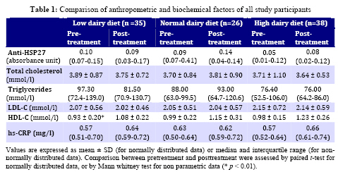
Iranian Journal of Pediatrics, Vol. 19, No. 1, March, 2009, pp. 41-46 Effect of a High Dairy Diet on Serum Antibody Titers to Heat Shock Protein 27 in Overweight and Obese Children Mohammad Safarian1, PhD; Rahim Vakili2, MD; PhD; Amirhossein Sahebkar3, PharmD; Mohsen Nematy1, PhD; Monireh Dahri3, BSc; Shima Tavallaie3, MSc; Elham Lotfian3, BSc; Mona Khorashadizadeh3, BSc; Gordon Ferns4, PhD; Majid Ghayour-Mobarhan*1,3, PhD 1. Department of Nutrition and Biochemistry, Faculty of Medicine, Mashhad University of Medical Sciences, Mashhad, IR Iran Received: 27/08/08; Revised: 23/09/08; Accepted: 25/11/08 Code Number: pe09006 Abstract Objective: An immune response to heat shock proteins appears to be involved in atherogenesis. To date, there has been no report on the impact of dairy or calcium consumptionon serum antibody titers to heat shock protein 27 (anti-HSP27).We have investigated whether an increase in dairy food consumption is capable of affecting serum antibody titers to heat shock protein 27 (anti-HSP27) level in children. Key Words: Dairy; Body Mass Index; Overweight; Obesity; Anti-HSP27; Calcium Introduction Heat shock proteins (HSPs) are a class of functionally related proteins whose expression is dramatically enhanced in response to various environmental stresses such as increases in temperature[1, 2]. This heat shock response is highly conserved through evolution[3,4]. HSPs function primarily as molecular chaperones. They play an important role in facilitating the folding of other cellular proteins, assisting the refolding of partially denatured proteins, preventing protein aggregation and targeting improperly folded proteins to specific degradative pathways[3,5,6]. Clinical studies have reported positive associations between plasma antibody titers to heat shock proteins (HSPs) and cardiovascular disease[7,8]. According to our previous studies, antibody titers to several HSPs are elevated in obese subjects without established coronary heart disease[9]. HSP27 is a member of small HSP family of proteins. In addition to its role as a chaperone, it has also been reported to have many additional functions including thermotolerance, inhibition of apoptosis, regulation of cell development and cell differentiation and signal transduction[10-14]. Recently it has been suggested that HSP27 may prevent obesity-induced insulin resistance possibly through suppression of TNF-mediated activation of IkappaB kinase-beta (IKK-beta)[15]. Previously it was shown that calcium ions control the mechanisms involved in the antibody formation by an antagonistic action on cell proliferation and cell differentiation which take place simultaneously in the immune response[16], and this interaction is probably due to the effect of calcium ions on the action of cyclic AMP and cyclic GMP[17,18]. However, to date, there has not been any report about the impact of calcium/dairy consumption on serum antibody titers to any of the heat shock proteins. In the present study,we wished to investigate whether or not serum antibody titers to HSP27 are influenced by increasing dairy consumption in overweight and obese children, who were placed on a calorie restricted diet. Subjects and Methods Subjects: Ninety-nine overweight or obese children were recruitedto participate in the study. Inclusion criteria were: An age between 12 and 18 years, body mass index (BMI) between 27 and 40 kg/m2 and a low-calcium diet (current calcium consumption500 mg/d and current dairy product consumption2 serving/d,as determined by food frequency and diet history). Participants were randomized toone of the following outpatient dietary regimens for 12 weeks:1) a calorie restricted, low-calcium diet providing a 500 kcal/d deficit from total energy expenditure and twoservings of dairy products/day (n=38); 2) a calorie restricted diet similar to that of group 1 and threeservings of dairy products/day (n=26); or 3) a calorie restricted diet similar to that of groups 1 and 2 and fourservings of dairy products/ day (n=35).Each serving was a moderate fat diary serving and consisted of 8 g protein, 300 mg calcium, and 5 g fat. Although the diets were individualized to achieve a500 kcal/person per day deficit, comparable advice was given to patients in all treatment groups, and diets were monitored weekly. All assessments were performed at baseline and at the end of the study after 12 weeks. Collection of serum samples: Blood samples were collected in the morning from each subject after an overnight fast. After being allowed to clot, the blood was then centrifuged at 2500 rpm for 15 minutes at room temperature to obtain serum. Hemolyzed samples were excluded from analysis. Serum was stored at -80⁰C prior to analysis. Routine biochemical analysis: A full fasted lipid profile comprising total cholesterol, triglycerides, low-density lipoprotein (LDL-C) and high-density lipoprotein cholesterol (HDL-C) was determined for each patient. Serum lipid concentrations were measured by routine enzymatic methods. hs-CRP was measured by a PEG-enhanced immuneturbidimetry method with an Alycon analyzer (ABBOTT, Chicago, IL, USA). Serum anti-HSP27 titers: Serum HSP27 antibody titers were measured using an in-house enzyme-linked immunosorbent assay (ELISA) as previously described[19]. Serum HSP27 antibody titers were measured using an in-house ELISA assay. Microtiter plates (Nunc Maxisorp, Nunc) were coated with 100ng per well recombinant human HSP27 dissolved in 50 ml carbonate buffer pH= 9.6 incubated for 18 hours at 4°C under humidified conditions. The wells were washed 3 times in wash buffer (PBS containing 0.05% Tween-20). Non-specific binding was reduced by blocking each well with 2% goat serum in PBS and 250 ml added to each well and incubated for 30 minutes in 37°C and 30 minutes at room temperature. Wells were washed 3 times with PBS. Serum was diluted 1:100 with 2% goat serum in PBS and 100ml added to each well in duplicated and incubated for 30 min at room temperature. After washing (4 times in wash buffer and 2 times in PBS), 100ml peroxidase conjugated-goat anti-human IgG (Sigma Sigma-Aldrich, INC., USA) diluted 1:500 with 2% goat serum in PBS, was added to each well, and incubated for 30 min at room temperature. After washing (4 times in wash buffer and 2 times in PBS), 100ml of TMB substrate (100ml of 6 mg/ml TMB in DMSO was added to 10 ml of 50 mM acetate buffer pH 4.5 containing 3 ml H2O2) was added per well and plate incubated for 15 minutes in the dark at room temperature. The reaction was terminated by adding 50ml 2M HCl per well. Optical density at 450 nm was measured using a Labsystems iEMS Reader MF Microtiter plate reader with a reference wavelength of 620 or 570 nm. The results were expressed in optical density units. Statistical analysis: All data were analyzed by using the statistical package for social sciences (SPSS v.11.5) software and were summarized and expressed as mean (±SD), or in the case of non-normally distributed data, as median and inter-quartile range. Statistical analysis of data was performed by paired t-test and a P-value less than 0.05 was considered significant. Findings Effect of dairy consumption on lipid profile: A comparison between lipid profile status at baseline and at the end of study showed that lipid profile was unaltered in all groups compared to the baseline levels values (P>0.05, Table 1), the only exception was the HDL-cholesterol concentration in the low-dairy diet group which was significantly increased at the end of the study (P<0.01, Table 1). Effect of dairy consumption on hs-CRP: Serum hs-CRP did not change significantly in any of the groups compared to the baseline levels (P>0.05, Table 1). Effect of dairy consumption on serum anti-HSP27: Serum anti-HSP27 titers in the subjects with the low-dairy diet was almost unaltered at the end of study (P>0.05, Table 1). In the subgroups of normal- and high-dairy diet, although serum anti-HSP27 level was slightly elevated, these changes did not reach statistical significance (P>0.05, Table 1). Discussion To our knowledge, this is the first report on the impact of calcium-rich diet on serum anti-HSP27. It has been reported that heat shock proteins 27 and 72 have the potential to prevent the activation of IKK-beta and JNK, respectively and this suggests that induction of heat shock proteins may blunt the adverse impact of obesity and fat overexposure on insulin function and thereby preventing obesity-induced insulin resistance[15,20]. Indeed, bimoclomol – a heat shock protein co-inducer being developed for treatment of diabetic neuropathy – and lipoic acid – suspected to be a heat shock protein inducer – have each demonstrated favorable effects on the insulin sensitivity of obese rodents, and parenteral lipoic acid is reported to improve the insulin sensitivity of type 2 diabetics[15]. In the present study serum anti-HSP27 was reduced in the low dairy-diet group and elevated in the normal- and high-dairy diet groups but all of these changes were less than significance level. The slight elevation in anti-HSP27 level with increasing dairy consumption could be attributed to the dairy-induced increase in free intracellular calcium because free intracellular calcium concentration appears to play a significant role in the induction of HSP gene expression[21], probably through activation of heat shock transcription factor[22,23]. This possible mechanism is supported by the result of two studies in which increasing the intracellular calcium concentration led to the induction of HSP gene expression in primary cultures of rat hepatocytes and human monocytic line U-937[24,25]. However, it is worth noting that extracellular calcium may also have an effect in the induction of HSP expression. In the study of Silomon et al, increase in extracellular calcium concentration was responsible for the [Ca2+] flux from the extracellular medium to the cytoplasm and therefore increasing the intracellular calcium concentrations and this mechanism is compliant with the finding of another study in which increasing extracellular calcium concentration augmented the expression of HSP60 and induced the expression of HSP72 in normal human keratinocytes cultured in serum free medium[26]. In our study, we did not find any significant change due to dairy calcium intakeon lipid profile confirming findings of previous studies[27,28]. The only significant change was detectedin HDL-C levels among the low-dairy diet group, as was shown before[29,30,31]. However it remains to be clarified that this significant change in HDL-C is due to the calcium intake or weight loss. As a limitation, it is worth noting that assessment of compliance with the intervention in this study was based on self-reporteddietary records which may not be an accurate indicator of true energy and dairy intake. However, we tried to overcome this limitation by a high quality of follow-up during the trial. Besides, the duration of follow-up in our study was rather short. The direction of changes in serum anti-HSP 27 suggests that over a longer period of time and with a largernumber of subjects a reliable effect might be observed. Conclusion On the basis of the present study, high dairy calcium consumption does not have any remarkable effect on the immune response to the HSP27 and it seems that factors other than dietary calcium are responsible for the modulation of HSP27 expression and the immunity toward this protein. Therefore, this finding may give insight for future research that are designed to identify micronutrients and dietary factors which are responsible for the regulation of serum antibody titers to heat shock proteins. Acknowledgment This research project has been financially supported by the Mashhad University of Medical Sciences Research Council and was approved by the MUMS Ethics Committee. The informed consentwas obtained from all participants. We express appreciation to the staff of the Nutrition Department of the Ghaem Hospital. The participation of the staff of the Bu-Ali (Avicenna) Research Institute of the MUMS is gratefully acknowledged. References
© 2009 by Center of Excellence for Pediatrics, Children’s Medical Center, Tehran University of Medical Sciences,All rights reserved. The following images related to this document are available:Photo images[pe09006t1.jpg] |
| |||||||||