 |
| About Bioline | All Journals | Testimonials | Membership | News |
|
||||||
|
||||||
Indian Journal of Pharmacology, Vol. 37, No. 5, September-October, 2005, pp. 304-308 Research Paper Antifungal activities of a steroid from Pallavicinia lyellii, a liverwort Subhisha S, Subramoniam A Tropical Botanic Garden and Research Institute, Pacha-Palode-695 562, Trivandrum District, Kerala Date of Submission: 30-Apr-2005 Code Number: ph05079 Abstract Objective: To determine the in vitro antifungal activity of Pallavicinia lyellii , a liverwort and to obtain clues about the active principle(s) and toxicity, if any.Materials and Methods: The in vitro antifungal activity of P. lyellii was studied against four test fungi ( A. niger, A. fumigatus, F. oxysporum and C. albicans ) using disc diffusion and direct dilution methods. Water, alcohol, and hexane extracts of P. lyellii were tested and the most active alcohol extract was subjected to sequential solvent fractionation. The promising hexane fraction was subjected to thin layer chromatography on silica gel and each spot on the gel was tested for activity and the active spot was chemically analyzed. The alcohol extract was evaluated for its short-term toxicity in mice. Results: Water, alcohol, and hexane extracts of P. lyellii showed varying levels of activity against the test fungi; the alcohol extract exhibited maximum activity. Out of the 4 test fungi , A. fumigatus was found to be the most sensitive . The alcohol extract was devoid of conspicuous short-term toxicity to mice. An active hexane fraction was separated from alcohol extract and from this fraction a steroid component with remarkable antifungal activity was isolated using thin layer chromatography (TLC). Conclusion: From P. lyellii a steroidal fraction with remarkable in vitro antifungal activity has been isolated. Further, the extract is devoid of conspicuous toxicity based on short-term toxicity evaluation in mice. Keywords: Antifungal agents, A. fumigatus , toxicity Introduction One of the lower groups of plants with tremendous potential for antifungal drug development is bryophytes. Bryophytes are closely linked with civilization, culture, beliefs, and ethics of humankind.[1] These organisms are also used in the ethnomedical field from times immemorial in many parts of the world.[1], [2] Bryophytes are used by different cultural groups for cuts, wounds and skin diseases suggesting that they protect the skin and open wounds from microbial pathogens.[2] Extracts of many bryophytes have been shown to possess varying levels of antibacterial and anticancer activities in vitro [2],[3],[4],[5] and many chemical constituents were isolated from bryophytes.[4], [6] The bryophytes form an important component of the forest ecosystem in India.[7] A recent exploration yielded about 250 species of bryophytes in Kerala state alone.[8] Although there is ample reason to believe that these plants could contain astonishing antimicrobial compounds, this is largely unexplored against infectious diseases. A literature search revealed no studies on the antifungal activity of Indian bryophytes. Studies done in other countries indicate that bryophytes are a rich source of antifungal agents. Cinnamolide from Porella and Makinoa showed activity against a few species of fungal dermatophytes.[2] Lunularin and lunularic acid isolated from Lunularia cruciata showed activity against many species of fungi such as Alternaria brassicola, Botrytis cinera, Septoria nodorum and Uromyces fabae .[2], [9] Dumortiera hirsuta , Sphagnum portoricense, and Orthotrichum rupestre were found to be active against Candida albicans . [2], [9] Antifungal compounds were isolated from the New Zealand liverwort Plagiochila faciculata .[10], [11] Out of several species of liverworts selected on the basis of traditional use or observations and screened for in vitro antifungal activities by the authors, Pallavicinia lyellii showed promising preliminary results. Therefore, a detailed study was carried out on the antifungal activity of this plant. Materials and Methods Plant materials Chemicals and reagents Preparation of water extract Alcohol extract Hexane extract Assay for antifungal activities Sabouraud (maltose) Agar (HI-MEDIA) was used as the medium for the antifungal assay by the disc diffusion method. Spread plates were prepared with the proper concentration of inocula. A known concentration of the extract in Tris - buffer (for water extract) or in 10% DMSO - Tris buffer (for alcohol extract) (40 µl) was added on each disc. Amphotericin B (Sigma), 0.25 mg/ml, and ketoconazole were used as a positive control and DMSO-Tris buffer (1:9) as a negative control for alcohol extract and only Tris-buffer was used for water extract. After 48-72 h of incubation at 27-28 oC, the inhibition zones from the centre of the disc to the inner margin of the surrounding fungal growth was measured in millimeters and recorded. Assay for antibacterial activities
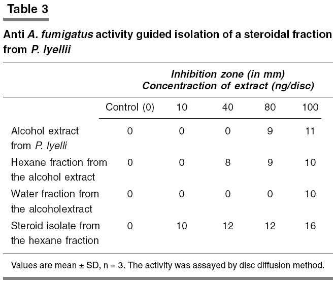
Chemical analysis of the active extract Toxicity evaluation in mice The behavior of the animals was observed daily for 1 h in the forenoon (10 to 11 A.M) for 14 days. The behavioral parameters observed were convulsion, grooming, hyperactivity, sedation, loss of righting reflex, and increased respiration. Initial and final body weights, water and food in take, and state of stool were observed. Rectal temperature was also recorded. The animals were killed on the fifteenth day. Hematological and serum biochemical parameters were determined. Liver, kidneys, spleen, and heart were dissected out, and weighed and observed for morphological and pathological changes. Hemoglobin was measured using hemoglobinometer with comparison standards. Glutamate pyruvate transaminase (GPT) and glutamate oxaloacetate transaminase (GOT) were measured by the method of Reitman and Frankel[15] and alkaline phosphatase by determining hydrolyzed phenol with antipyrine.[16] The peritoneal macrophage and total leucocyte count were done.[17] Statistical analysis Results The antifungal activity of different extracts of P. lyellii against A. niger , A. fumigatus, F. oxysporum, and C. albicans are given in [Table - 1]. Although all the extracts showed varying levels of activity against all the test fungi, the alcohol extract was found to be more active than water and hexane extracts. Out of the four test fungi, A. fumigatus was found to be more susceptible. The growth of this organism was inhibited at a concentration as low as 80 ng/disc of alcohol extract, whereas the growth of F. oxysporum , A. niger, and C. albicans was inhibited at 10, 50 and 100 µg/disc, respectively [Table - 1] and [Table - 3] The inhibition of growth of A. fumigatus by the alcohol extract was confirmed by directly adding into the liquid medium. As shown in [Table - 2], the growth inhibition was observed over a wide range of concentrations. The mycelia growth was inhibited more than 50% at 1 µg/ml; the inhibition was almost complete at 1 mg/ml level. Thus, it was found to be more potent than the commonly used antifungal agent, ketoconazole, which showed 82% growth inhibition at 1 mg/ml. In contrast to the antifungal activity, the extract showed very marginal antibacterial activity. In the case of E. coli, the extract showed significant inhibition zone in the disc diffusion assay at 250 µg /disc, while S. aureus was found to be resistant to this extract even at a high concentration of 10 mg/disc [Table - 4]. When the alcohol extract was further fractionated by sequential solvent extraction, the activity was found in the hexane and water fractions [Table - 3]. In the case of the hexane fraction of alcohol extract, concentration that was required for measurable inhibition of A. fumigatus growth in the disc diffusion assay was approximately 40 ng/disc as against 80 ng/disc (2.0 µg/ml; 40 µl/disc) of the original alcohol extract. The yield of the ethanol extract was 7.5% of the dry plant powder, while the yield of the hexane fraction was 65% of the extract. The chloroform, ethyl acetate, and butanol fractions were inactive even at 100 ng/disc level, while water fraction showed activity at 100 ng/disc. The yield of this water fraction is only 7% of alcohol extract. Upon chemical analysis, the water fraction showed the presence of coumarins. When the hexane extract was subjected to TLC separation on silica gel using different solvent systems [hexane-ethyl acetate (1:1), chloroform-methanol (8:2) and chloroform], the solvent chloroform was found to give better separation [Figure - 1]. The extract was resolved into several spots. The chromatograms were sprayed with different reagents or exposed to UV and inspected.[14] Each spot in preparative TLC was identified on the basis of relative mobility, and scrapped off and eluted with chloroform and tested for anti A. fumigatus activity using disc diffusion method. The fast moving dominant band/spot was found to be very active. This isolate showed activity at 10 ng/disc. As judged from Lieberman′s reaction, this isolate was a steroid [Figure - 1]. The yield of the steroid isolate was 34% of the hexane fraction. In short-term toxicity evaluation, none of the parameters studied was influenced by the alcohol extract administration for 15 days except serum triglyceride level which was significantly decreased at the high dose (500 mg/kg) [Table - 5]. Discussion This study reports for the fist time the potent antifungal activity of P. lyellii . The active fraction obtained from this liverwort showed varying levels of activity against all the four test fungi. This suggests that it has a broad spectrum of activity, although the degree of susceptibility could differ between different organisms. There is a need to test the in vivo activity of the extract apart from the effect on many other fungi. It is of interest to note that the plant extract appears to be nontoxic as judged from the general short-term toxicity study in mice. The limited studies clearly indicate that the active principle is a steroid. Further studies are under progress in this laboratory to characterize the active principle and the mechanism of action. The drug inhibits the germination of the spore, as well as the multiplication of mycelia. The active principle, being a lipophilic steroid is likely to act intracellularly. P. lyellii can be easily obtained through cultivation. The plant material appears to be an attractive material for antifungal drug development. There is an urgent need to develop new antifungal agents. Almost all of the antifungal agents, which are currently in use are relatively expensive and have toxic side effects.[18] Acknowledgments The authors acknowledge Dr. G.M. Nair, Director, Tropical Botanic Garden and Research Institute (TBGRI), and late Dr. G. Sreekandan Nair, former Director, TBGRI for their keen interest and encouragement in the course of this research work. Dr. C.K.C.S. Pillai, Professor of Botany (retired), MSM College, Kayamkulam, Kerala State, Dr. P.V. Madhusoodanan, Professor of Botany, University of Calicut and CN Manju, Research Scholar, University of Calicut are gratefully acknowledged for their help in the identification of the plant species. The authors express their sincere thanks to Mr. G. Santhoskumar, Animal House Technician, for providing technical assistance in the execution of animal experiments. References
Copyright 2005 - Indian Journal of Pharmacology The following images related to this document are available:Photo images[ph05079t3.jpg] [ph05079t5.jpg] [ph05079f1.jpg] [ph05079t2.jpg] [ph05079t1.jpg] [ph05079t4.jpg] |
| |||||||||
![[Table - 1]](/showimage?ph/photo/ph05079t1.jpg)
![[Table - 2]](/showimage?ph/photo/ph05079t2.jpg)
![[Table - 4]](/showimage?ph/photo/ph05079t4.jpg)
![[Figure - 1]](/showimage?ph/photo/ph05079f1.jpg)
![[Table - 5]](/showimage?ph/photo/ph05079t5.jpg)